
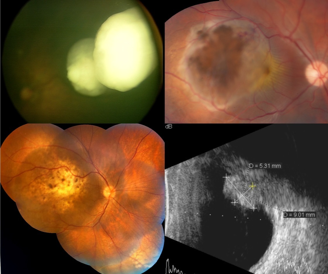
Intraocular tumors include primary tumors arising within the eye and secondary tumors that spread from another site. Early detection, multimodal imaging and coordinated management are important for protecting life, preserving the eye and retaining useful vision where possible.
Ocuprime provides diagnostic evaluation and evidence-based management led by ocular oncologist Dr. Sonal Yadav.
At a glance
Conditions and treatments
- ✓Retinoblastoma
- ✓Choroidal melanoma
- ✓Choroidal metastasis
- ✓Choroidal hemangioma
- ✓Choroidal granuloma
- ✓Optic nerve head melanocytoma
- ✓Retinal pigment epithelial tumors
Retinoblastoma
Dedicated retinoblastoma programme →
Read Ocuprime's parent-focused guide to warning signs, examination, treatment, genetic counselling and follow-up.
Overview
The most common primary intraocular malignancy of childhood, often presenting with a white pupillary reflex or strabismus.
Evaluation
Examination under anesthesia, fundus photography, ultrasonography and MRI support classification and treatment planning.
Treatment
Care may combine systemic or targeted chemotherapy with focal laser, cryotherapy or thermotherapy. Advanced, non-salvageable eyes may require enucleation followed by implant and prosthetic rehabilitation.
Choroidal melanoma
Overview
The most common primary intraocular malignancy in adults, arising from melanocytes in the uveal tract.
Evaluation and treatment
Ultrasound, autofluorescence and enhanced-depth OCT define the lesion and risk features. Treatment planning may include plaque radiotherapy co-management, thermotherapy or enucleation, together with systemic surveillance.
Choroidal metastasis
Overview
Secondary tumors in the choroid most often originate from systemic cancer, including breast or lung cancer.
Our approach
Multimodal imaging and, when necessary, fine-needle aspiration assist diagnosis. Treatment is coordinated with oncology and may involve systemic therapy or radiotherapy.
Choroidal hemangioma
Overview
A benign vascular tumor that may impair vision through subretinal fluid or macular edema.
Our approach
Angiography, OCT and ultrasound guide observation or treatment such as photodynamic therapy, thermotherapy or radiotherapy in selected cases.
Choroidal granuloma
Overview
Inflammatory or infectious lesions associated with conditions such as tuberculosis or sarcoidosis can mimic a tumor.
Our approach
Imaging and systemic investigation identify the cause so antimicrobial or immunomodulatory treatment can be directed appropriately.
Optic nerve head melanocytoma
Overview
A usually benign, deeply pigmented lesion of the optic disc that can rarely grow or cause local complications.
Our approach
Baseline photography, OCT and ultrasonography allow long-term surveillance and help distinguish stability from transformation.
Retinal pigment epithelial tumors
Overview
Rare lesions range from benign congenital hypertrophy or adenoma to malignant adenocarcinoma.
Our approach
OCT, autofluorescence and angiography support diagnosis. Stable benign lesions may be observed; progressive lesions are considered for local treatment or surgery.
Academic references and authored works
- Yadav SP, Tomar AS, Finger PT, Gallie B. Local Chemotherapy for Intraocular Tumors. In Surgical Ophthalmic Oncology: A Collaborative Open Access Reference. ↗
- Yadav SP (Editor). Retinoblastoma: Diagnosis to Management. Nova Science Publishers. ↗
- Yadav SP, Tomar AS, Finger PT. Topical Chemotherapy for Ocular Tumors. In Surgical Ophthalmic Oncology.