Dedicated care for children and families
Retinoblastoma Treatment
Stage-informed, multidisciplinary care for childhood retinoblastoma, with life, eye preservation and useful vision considered in that order.
Director & Chief Surgeon · Ophthalmic Plastic Surgeon, Orbit Specialist & Ocular Oncologist
Retinoblastoma is a rare cancer of the retina that occurs mainly in infants and young children. A white glow in the pupil or a new squint may be the first sign noticed by a parent, often in a photograph.
At Ocuprime Eye Institute, retinoblastoma care is organized around accurate staging, timely treatment, collaboration with pediatric cancer specialists and clear support for families throughout treatment and follow-up.
From Dr Sonal“A retinoblastoma diagnosis is frightening for any family. Our first priority is the child's life, followed by saving the eye safely and preserving useful vision wherever possible.”
Early signs parents and pediatricians should not ignore
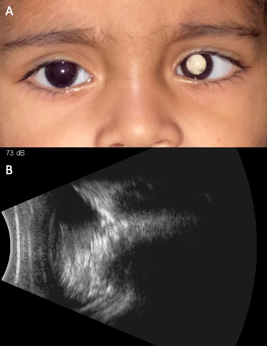
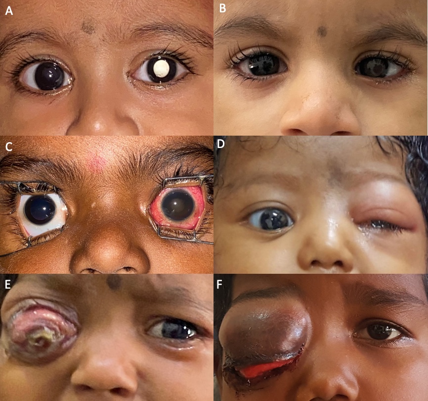
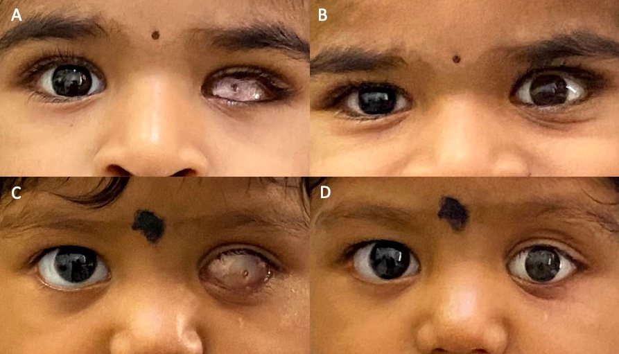
The clinical images below illustrate presenting signs, including advanced disease. The symptom descriptions explain what parents and pediatricians should look for.
White pupil reflex (leukocoria)
A white or pale glow in the pupil, especially in flash photographs or dim light, is the most common presenting sign and needs prompt examination.
New squint (strabismus)
A newly inward- or outward-turning eye is another important presentation and should not be assumed to be harmless.
Other changes
Persistent redness, swelling, pain, reduced vision or an unexplained change in a young child's visual behavior also requires assessment.
Our treatment philosophy: life, eye and vision
Protect the child's life
The first priority is control of the cancer and prevention or treatment of spread beyond the eye.
Preserve the eye when safe
Eye-conserving treatment is considered according to tumor group, laterality, location, response and the safety of continued treatment.
Preserve useful vision
When tumor location and treatment safety allow, therapy is planned to reduce damage to the retina, macula, optic nerve and other healthy structures.
Diagnosis, imaging and staging
Examination under anesthesia
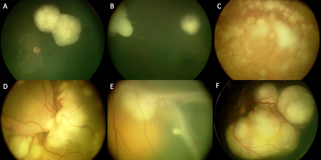
Young children commonly require a detailed examination under anesthesia so both eyes can be mapped accurately with retinal photography and indirect ophthalmoscopy.
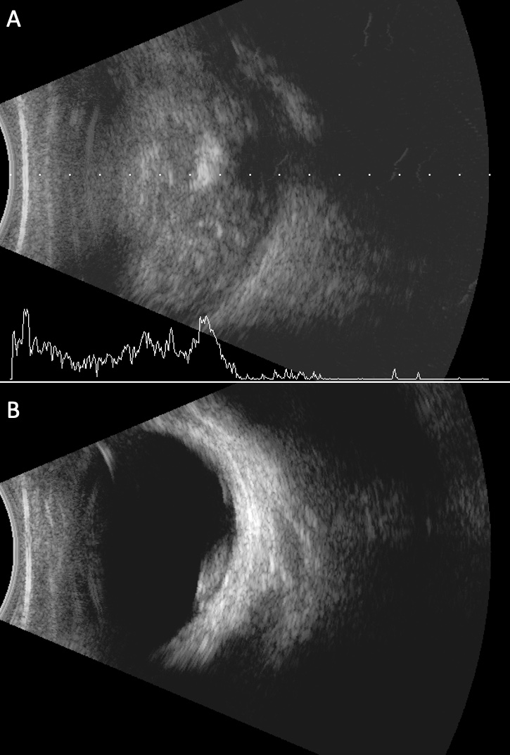
Ocular imaging
Ultrasonography and retinal imaging help document the tumor, calcification, retinal detachment and treatment response.
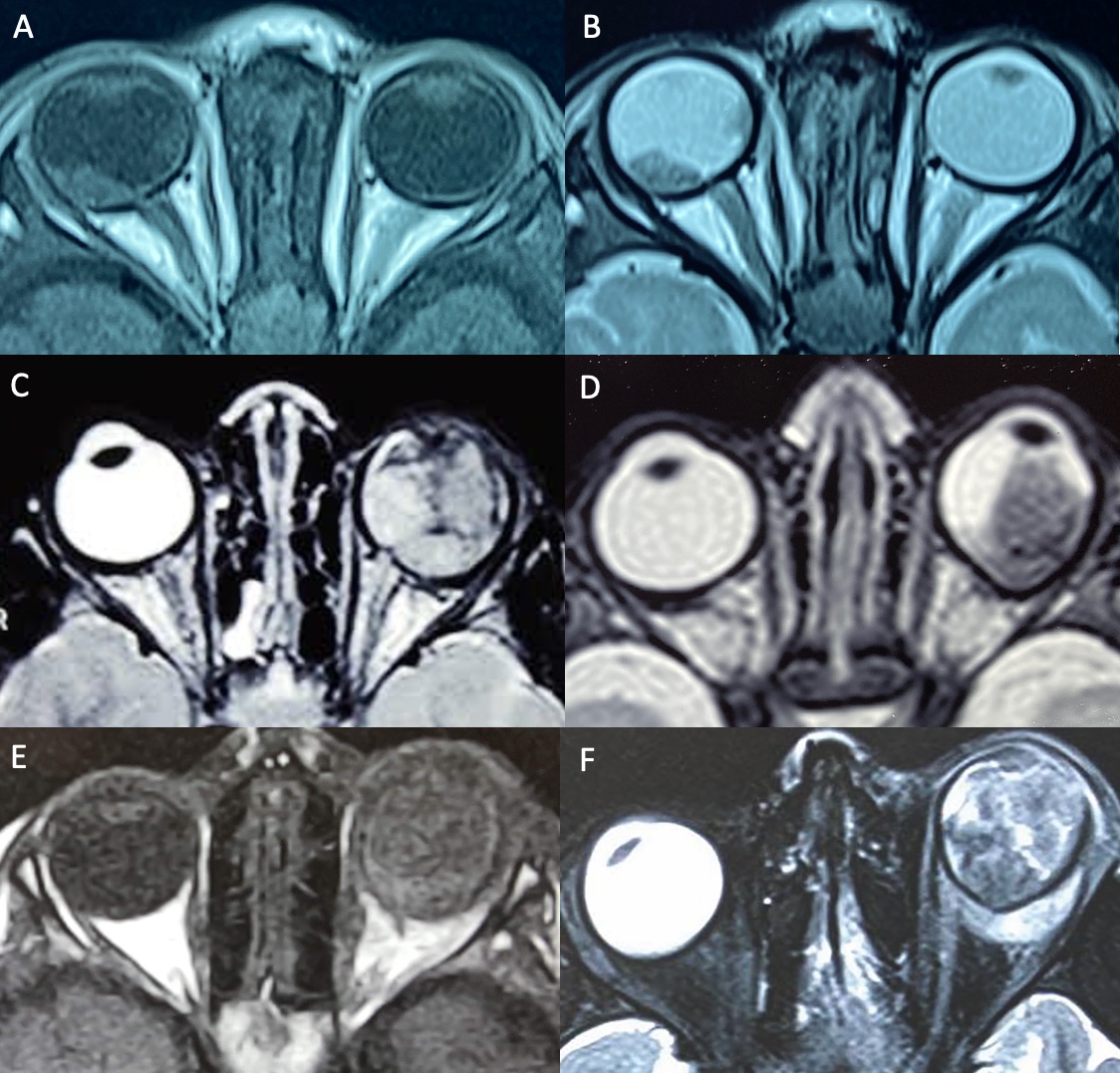
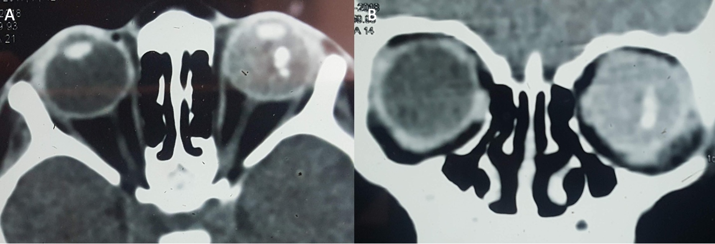
MRI and systemic assessment
MRI is used when indicated to assess the optic nerve, orbit and brain. Additional investigations depend on the clinical stage and multidisciplinary recommendation.
Classification
Treatment is individualized using the extent of disease in each eye, whether one or both eyes are involved, visual potential and possible hereditary RB1-related disease.
Chemotherapy and focal treatment
Systemic chemotherapy
Intravenous medicines can shrink tumors and treat disease risk beyond an individual eye; local treatment is then used to consolidate control when appropriate.
Intra-arterial chemotherapy
At suitably equipped specialist centers, chemotherapy can be delivered through the ophthalmic artery to selected eyes. Suitability depends on age, anatomy, tumor group and team assessment.
Intravitreal chemotherapy
Medicine injected into the vitreous may be used for selected vitreous seeds or recurrent intraocular disease using established safety techniques.
Laser, thermotherapy and cryotherapy
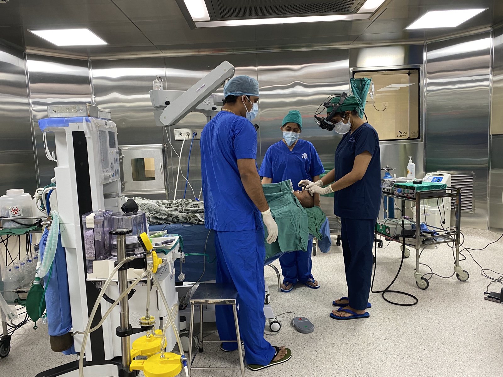
Focal treatments destroy smaller tumors or residual disease and are selected according to tumor size and retinal location.
When eye removal is necessary
Enucleation
A large or high-risk tumor may make eye removal the safest way to protect the child's life. Continuing eye-salvage treatment is not appropriate when cancer control is uncertain.
Pathology-guided next steps
The removed eye is examined for high-risk features that may influence whether additional chemotherapy is recommended.
Prosthetic rehabilitation
A primary orbital implant and later custom artificial-eye fitting help restore orbital volume, eyelid support and a natural appearance.
A multidisciplinary programme
Coordinated specialists
Depending on need, the care team includes ocular and pediatric oncologists, pediatric anesthesiologists, interventional specialists, neuroradiologists, pathologists and radiation oncologists.
Genetic counselling
Bilateral, multifocal or very early retinoblastoma may indicate a heritable RB1 change. Genetic counselling helps families understand testing, sibling screening and future surveillance.
Follow-up and survivorship
Repeated examinations monitor regression and recurrence. Long-term follow-up also considers the fellow eye, vision development, treatment effects, prosthetic care and hereditary cancer risk.
Family support
The programme emphasizes understandable explanations, written treatment plans and practical guidance throughout diagnosis, treatment and recovery.
Clinical and academic leadership
Focused training
Dr. Sonal Yadav trained in ocular oncology in India and the United States, including at The New York Eye Cancer Center, with observer training at MD Anderson Cancer Center.
Retinoblastoma reference work
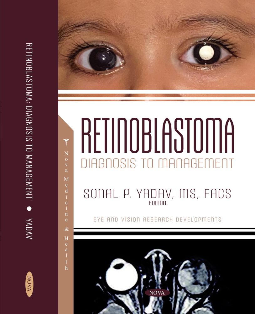
As editor of Retinoblastoma: Diagnosis to Management, she worked with international contributors on diagnosis, treatment and the global context of this childhood eye cancer.
Individual treatment decisions
No single treatment sequence suits every child. Recommendations follow examination findings, tumor group, genetic context, response and multidisciplinary review.
Authoritative patient information
These external sources support the general medical information on this page. Individual treatment recommendations require a clinical examination and multidisciplinary review.
Speak with our team